96 Hour Chick Embryo Serial Section
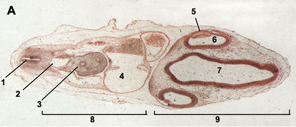
4 Day (96 hour) Chick Embryo. Extending out from the embryo is a thin delicate membrane with blood vessels at its surface. This membrane, called the allantois, expands as the embryo develops until it completely lines the shell.The transparent sac surrounding the embryo is the amnion.The material in the amnion is principally water from the egg white.
This membrane is made up of a bladderlike median ventral diverticulum of the hindgut endoderm, covered with splanchnic mesoderm. Acid pro download. It connects with the hindgut, which will be found only in sections posterior to the posterior intestinal portal at this stage of development. Ultimately, this double membrane will fill the exocoel, and its outer layer of mesoderm will fuse with mesoderm of the chorion and the aminion and finally with the splanchnic mesoderm of the yolk sac splanchnopleure. Its function in the chick is related to respiration and excretion. The entire outer covering of the chick embryo is of ectodermal origin and is made up largely of squamous epithelium but will later also include horny scales, feather germs, quills and barbs, claws, beak coverings, and a temporary eggtooth.
By evaginations from the surface, the linings of the following structures are also derived from ectoderm: the mouth (stomodeal portion and stomodeal hypophysis); cloaca (proctodeal portion); visceral clefts (peripheral halves); nostrils; eye chamber and lens; otic vesicles; and external auditory meatus.